Scientific Program

Conference Series Ltd invites all the participants across the globe to attend 3rd International Conference & Exhibition on Tissue Preservation and Biobanking (10 Plenary Forums - 1 Event)
San Francisco, California, USA.

Day 2 :

Keynote Forum

Aubrey De Grey

SENS Research Foundation, USA

Keynote: Cryopreservation of organs and organisms: Signs of a new era

Time : 09:00-09:25

Conference Series Biobanking 2017 International Conference Keynote Speaker Aubrey De Grey photo
Biography:

Aubrey De Grey is a Biomedical Gerontologist based in Mountain View, California, USA, and is the Chief Science Officer of SENS Research Foundation, a California-based 501(c) (3) biomedical research charity that performs and funds laboratory research dedicated to combating the aging process. He received his BA in
Computer Science and Ph.D in Biology from the University of Cambridge. His research interests encompass the characterization of all the types of self-inflicted cellular and molecular damage that constitute mammalian aging and the design of interventions to repair and/or obviate that damage. He is a fellow of both the
Gerontological Society of America and the American Aging Association, and sits on the Editorial and Scientific Advisory Boards of numerous journals and organizations. He is a highly sought-after speaker who gives 40-50 invited talks per year at scientific conferences, universities, companies in areas ranging from pharma to life insurance, and to the public.

Abstract:

Huge numbers of people worldwide die while on waiting lists for organ transplantation, purely because no one sufficiently

immunocompatible dies sufficiently nearby. This tragedy would be substantially reduced if organs could be maintained in a viable state long enough to survive intercontinental travel, and it would be virtually eliminated if organs could be preserved indefinitely. Moreover, in the research arena cryopreservation of biological material has the potential to eliminate huge manpower costs in the maintenance of breeding populations of laboratory animals (such as fruit flies) and to enhance the quality of tissue slices. I will review a number of dramatic recent advances in the cryopreservation field that promise to deliver immense progress in all these areas.

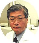
Conference Series Biobanking 2017 International Conference Keynote Speaker Yaegaki K photo
Biography:

Yaegaki K had received Doctor of Dental Science, and then completed his Ph.D in Medicine from Kureme University in Medical Biochemistry and Post-doctoral studies from University of British Columbia. He was trained as an Oral Maxillofacial Surgeon at School of Medicine Kurume University. His speciallity was facial injuaries. He is the Director and Head of Oral Health at Nippon Dental University, which is the oldest dental school in Asia, also a Dean of Ph.D Program. He has published more than 100 papers and 20 books.

Abstract:

We treated the liver cirrhosis by transplantation of hepatocyte-like-cell derived stem cells from human exfoliated deciduous
tooth (SHED) into the rata or swine with cirrhosis, NASH or bilious cirrhosis model. SHED at 3 passage was separated by
magnetic sorting with CD117 antibody. For hepatic differentiation, CD117+ SHED were grown in DMEM supplemented with
insulin-transferrin-selenium-x (ITS-x), embryo-tropic-factors(ETF) and hepatocyte-growth-factor (HGF) for 5days: IMDM
supplemented with ITS-x, ETF, HGF, dexamethasone and oncostatin for another 11 days. F344-Nude rats were employed
for this study. Carbon tetrachloride (CCl4) was administered by intra-peritoneal injection for 12 weeks to induce cirrhosis.
Hepatocyte-like-cells (2 x 10⁶ cells/ animal) suspended in Hank`s Balanced Salt Solution were transplanted into the spleen. The
vehicle was injected to the positive control group. Non-cirrhosis-models were used as negative control group. Animals were
sacrificed 4 weeks after the transplantation. Immunocytochemistry observation of the hepatically differentiated cells strongly
demonstrated positive staining for albumin, IGF-1, α-feto-protein, HNF4α and CPS-1. The histopathological analysis, HE and
Masson’s trichrome staining, indicated a significant decrease of fibrous tissue in the transplantation group with comparison to
the positive control group. Healthy liver tissues were recovered by the transplantation. Moreover, serological test results revealed
significant differences between the groups. Serum ALT levels of the test group dramatically decreased to one-third compared
to the positive control group. Activities of albumin, bilirubin, BUN, HA levels were also recovered. The xenotransplantation of
hepatocyte-like cells from human tooth transplanted into the liver with severe failure, demonstrated their capacity to preform
positively because of drastic decreasing fibrous tissues. Together, these findings suggested. Hepatocyte-like cells derived from
SHED are a potential source for treating chronic liver injuries such as cirrhosis.

Keynote Forum

Luiz C. Samapio

Texas Heart Institute, USA

Keynote: Maximizing cardiac repair: Should we focus on cells or matrix?

Time : 09:50-10:15
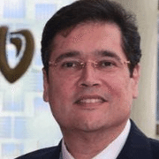
Conference Series Biobanking 2017 International Conference Keynote Speaker Luiz C. Samapio photo
Biography:

Cardiac extracellular matrix (ECM) is a complex architecturally-organized mixture of proteins glycosamino-, andproteoglycans that contains an embedded vascular network. Furthermore, ECM binds growth factors that can drive vasculogenesis. In fact, during cardiac development, cell-matrix interactions give rise to regional cell fate specifications including atrial ventricular, pacemaker, vascular, and neural cells. We hypothesize that cardiac ECM plays a critical role in SC fate, location and function after myocardial injury. Using decellularized ECM from neonatal, adult and old animals, we tested
the hypothesis that ECM composition and architecture provides physical, mechanical and biochemical cues that direct stem
or progenitor cell fate – and show evidence that age, sex and disease state can alter those biologic cues and in turn alter cell
phenotype. Furthermore, by exploring the gene expression profiles of SCs in response to infarcted heart matrix vs collagen
type 1 or non-infarcted matrix, we have begun to define cues that may begin to explain clinical outcomes after cell therapy.
We have also transplanted repopulated and “naked” ECM in vivo either as a cardiac patch after infarction or as a whole heart
to investigate SC recruitment and differentiation in response to acellular or recellularized ECM. Finally, we are convinced that
creating and being able to manipulate the right environment can be more valuable than engineering cells. Our perfusion-based
method to remove cells from myocardium giving rise to decellularized ECM (dECM) with intact micro and macrostructure,
and a patent vascular tree has been proved to be the perfect environment for stem cells.

Abstract:

Dr. Luiz C. Sampaio has devoted his entire career to reducing the burden of cardiovascular disease, first as a cardiovascular surgeon then as a regenerative medicine researcher. In his current positions as associate medical director for the Department of Regenerative Medicine Research and director of the Cullen Cardiovascular Research Laboratories at Texas Heart Institute, he is able to apply his medical and surgical skills to developing new approaches to preventing,
diagnosing, and treating heart disease.

  • Biobanking | Cryopreservation| Vitrification | Biorepository & Biospecimen| Stemcell | Regenerative medicine| Tissue engineering | Rejuvenation
Location: Plaza I

Chair

Heiko Zimmermann

Fraunhofer Institute for Biomedical Engineering, Germany

Co-Chair

Ken yaegaki

Nippon Dental University School of Life Dentistry, Japan

Session Introduction

Aijun Wan

University of California, USA

Title: Engineering stem cells and biomaterials to treat birth defects before birth

Time : 14:20-14:40

Speaker
Biography:

Aijun Wang is the Co-Director of the Surgical Bioengineering Laboratory and an Assistant Professor at the University of California Davis School of Medicine. His research interests center on Engineering Stem Cells and Biomaterials to develop novel regenerative medical therapies, especially surgical treatments for congenital anomalies. His lab has successfully combined Tissue-Engineering technologies with the most advanced fetal intervention, and developed novel biomaterial and stem cell-based treatments (including nanofibrous materials, fetal membrane, decellularized extracellular matrix, iPSC-derived stem cells, placenta-derived stem cells) for devastating birth defects, such as spina bifida. Currently, his team are performing IND enabling studies to move novel stem cell based therapies to clinical applications.

Abstract:

Statement of the Problem: Birth defects are common, affecting approximately 3% of all live births in the United States. Congenital defects can result in significant morbidity and are the most common cause of infant mortality, accounting for a minimum of 20% of all infant deaths worldwide. Improvements in prenatal screening and diagnostics- including ultrasonography, chorionic villus sampling, amniocentesis, fetal magnetic resonance imaging, and, most recently, circulating fetal DNA in maternal blood have allowed earlier diagnosis of a range of congenital disorders. With prenatal diagnosis came the pursuit of prenatal interventions to prevent or address pathology that developed in utero.
 
Methodology & Theoretical Orientation: Our lab has been developing fetal tissue engineering approaches using different types of stem cells and biomaterials to treat a variety of birth defects before birth. One example is in utero transplantation of stem cells with extracellular matrix-based scaffolds to treat spina bifida. Our recent data have shown that treatment with early
gestation placenta-derived mesenchymal stromal cells (PMSCs) during in utero repair cures spina bifida-associated motor function at birth in a fetal lamb model. Based on the previous studies, we are refining the treatments and pursuing IND enabling studies for future clinical translations. We are also developing in utero stem cell transplantation approaches to treat other birth defects such as hemophilia, tracheal occlusion and congenital diaphragmatic hernia.
 
Conclusion & Significance: Stem cell therapies have the potential for use in fetal Tissue Engineering applications, as a vehicle for fetal Genetic Engineering, for the induction of fetal tolerance for postnatal transplantation, and perhaps even for use in fetal treatment of potential adult disease. The fetal environment is ideal for the introduction of cell-based therapies prior to
development of pathology. As with any new therapeutic modality, the ideal technique, potential risks, and long-term efficacy need to be determined.

Elvira Famulari

Molecular Biotechnology Center, Italy

Title: Cell therapy for Crilger Najjar Type I syndrome

Time : 18:00-18:15

Biography:

Elvira Famulari is pursuing Ph.D in Molecular Biotechnology Center from University of Turin Italy.

Abstract:

Crigler-Najjar Syndrome type I (CNSI) is a recessive disorder caused by a rare mutation in Ugt1a1 gene that encodes for the enzyme Ugt1 involved in bilirubin metabolism. This mutation causes high levels of unconjugated bilirubin in blood, leading to brain damage and early lethality. CNSI patients are treated with extended daily phototherapy but the only effective therapy is liver transplantation. However liver transplantation is not exempt from complications, therefore hepatocyte-like cells derived from stem cells represent an interesting alternative. The goal of this project is to evaluate the use of human adult liver stem cells (HLSC) in treating Crigler-Najjar Syndrome type I (CNSI) in a model represented by Ugt1 deficient mice closely mimicking the pathological manifestations in CNSI patients. The Ugt1+/- mice have been backcrossed with NSG for 9 generations in order to derive these mice in a pure NSG background. By recellularising decellularised rat liver scaffolds with HLSCs ex vivo, we found that HLSC can differentiate in functional hepatocytes expressing Ugt1 protein as from 7 days differentiation. A significant improvement in survival of phototherapy-treated NSG/Ugt1a1-/- pups following injection of HLSC in the liver was observed. Immunohistochemically and immunofluorescence analyses show that HLSC can engraft in the liver of NSG/Ugt1a1-/- mice and express missing enzyme. HLSCs thus show great potential not only for the treatment of CNSI but also for most metabolic liver disorders.

Speaker
Biography:

Dr Wael Abo Elkheir has completed his Ph.D at the age of 35 years from Cairo University. He is the co-founder and board member of the Egyptian Society for Progenitor Stem Cell Research, a society initiated with the mission of enhancing scientific research and cooperation in the field of stem cell research and regenerative medicine. He is the director of a number of registered clinical trials in the field of stem cell therapy, especially for neuro-regeneration and musculoskeletal disorders. He has published more than 20 papers in reputed journals.

Abstract:

Background: Chondrogenesis is a well-orchestrated process in which chondroprogenitors undergo proliferation and chondrocyte differentiation. Because cartilage lacks blood supply, it shows poor regenerative power and subsequent wound healing. Cartilage degeneration caused by disease or trauma carries great clinical implication on the function of joints. The end stage of cartilage damage frequently leads to osteoarthritis (OA), resulting in a significant impairment of the quality of life of millions of people. MSCs are multilineage progenitors responsible for the normal turnover and repair of mesenchymal tissues, such as bone, cartilage, ligament, and fat. In-vitro expanded MSCs can differentiate into chondrogenic, adipogenic and
osteogenic lineages.
 
Aim: The objective of this work was to evaluate the regenerative effect of cell therapy in cases of acute and chronic OA canine models of surgically induced partial thickness chondral defects injected with autologous bone marrow derived MSCs.
 
Study Design: This work was done on 24 knees of male domestic mongrel dogs. OA was induced by doing surgical chondral defects then injected intra-articular with MSCs. Dogs were divided into the following groups: acute (injected after 1 day), chronic (after 1 month) and control group received conventional treatment. The dogs were sacrificed after 1, 2, 6 and 8 weeks
of injection. Assessment by histological scoring of cartilage repair (Os Score) for blind randomized samples and by clinical examination for lameness degree scores was done.
 
Results: Our results showed that dogs possess characteristics that are not found in traditional rodent models and confirmed the efficacy of direct intraarticular injection of MSCs to home and function in cartilage defects both in acute and chronic lesions.
 
Conclusion: This study concludes that the local delivery of MSC is a good therapeutic option for O A.

Speaker
Biography:

Hala Gabr is a renowned researcher in stem cell biology and therapy in Cairo University. She is the Director of the Pediatric Bone Marrow Transplantation and Cellular Therapy Lab in Cairo University. She is the Co-founder of the Egyptian Society for Progenitor Stem Cell Research, the leading stem cell research body in
Egypt. She has published more than 30 papers in reputed journals and is an Editorial Board Member of a number of reputed journals. She has supervised nearly a hundred Ph.D and Master thesis in stem cell research.

Abstract:

Liver transplantation represents the only definitive treatment for cases of end stage liver failure. However, this procedure is hindered by a number of obstacles, namely; the marked shortage of liver donors, major operative procedures and lifelong immune suppression, in addition to the high expenses. Regenerative medicine, based on cellular approach for repairing and replacing damaged tissues and organs, is a rapidly growing field of Medicine. Due to the heavy burden of liver diseases in Egypt, this study was designed to evaluate the efficacy of cellular therapy in the form of hepatocytes derived from patients' own haematopoietic stem cells (HSCs), transplanted directly through intra-splenic injections in patients with liver cirrhosis grade B and C Child-Turcotte-Pugh score (CTP B). 100 patients with liver cirrhosis CTP B score were divided into two groups according to the principle of treatment. Group (A) consisted of 50 patients (25 Child B and 25 Child C), who received hepatocytes derived from patients own (HSCs) in addition to conventional treatment. Group (B) received regular conventional treatment. Both groups of patients were followed up for six months after transplantation for assessment of liver functions. There was a significant improvement in the degrees of ascites, lower limb edema, HE, CTP scores and MELD scores in patients treated with hepatocytes derived from HSC. Also, we observed a slight improvement in serum albumin, prothrombin concentration and international normalized ratio in stem cell treated group. No procedure related complications were encountered. We demonstrated the safety and short term efficacy of autologous bone marrow derived hepatocyte transplantation for the support of cirrhotic liver.

Jin-Ye Wang

Shanghai Jiao Tong University, China

Title: Natural Polymer, Zein for Tissue Regeneration

Time : 17:00-17:20

Speaker
Biography:

Jin-Ye Wang has received her Ph.D from Tohoku University, Japan (1992). She was working as a Professor of Shanghai Institute of Organic Chemistry, Chinese Academy of Sciences (2000-2009), Adjunct Professor of Shanghai Jiao Tong University (2003-2009), and now Professor of Biomedical Engineering and Team Leader of Biomaterials Lab, Shanghai Jiao Tong University. She has published over 80 SCI papers, 4 books (chapters), and 13 Chinese patents and one US patent were authorized. She was the Invited Speaker of European Conference on Biomaterials, Pacifichem et al., awarded by the Hundred Talent Program of the Chinese Academy of Sciences (1999), Life Sciences Prize from Meiji Dairies Corporation (2008) et al. Her research interests include Tissue Engineering, Controlled Release and Fluorescent Probe, Biomimetic Materials and Biointerfaces
 

Abstract:

Zein is the major storage protein of corn and comprises 40-50% of total endosperm proteins. Zein has been used as microspheres to delay the release of drugs and to protect the drugs from degradation by pepsin, thus can release the
drugs for a long time. Our laboratory has developed zein as a novel and potential biomaterial for Tissue Engineering. Firstly, a three-dimensional zein porous scaffold was prepared and showed to be suitable for culture of various cell lines and primary cells such as human umbilical vein endothelial cells (HUVECs) and mesebchymal stem cells (MSCs) in vitro. The scaffolds are characterized with interconnected pore, controllable pore sizes, especially excellent mechanical properties, which are controllable and suitable to act as bone substitutes. Next, we examined its tissue compatibility in a rabbit subcutaneous implanting model, the histological analysis revealed a good tissue response and degradability. The third, zein porous scaffolds modified with fatty acids have shown great improvement in mechanical properties and also good cell compatibility in vitro. Besides, the complex of zein porous scaffold and mesenchymal stem cells (MSCs) could effectively promote the ectopic bone formation in nude mice and the repair of critical-sized bone defects in the rabbit model.

Shrikant L Kulkarni

Kulkarni Clinic, India

Title: Hydro-pressure therapy in chronic kidney diseases

Time : 16:40-17:00

Speaker
Biography:

Shrikant L Kulkarni has completed his MS (General Surgery) in 1975 from B J Medical College Pune, Maharashtra, India and his MBBS from Miraj Medical College,Sangli. Since 1971, he has worked at several Government Hospitals like the Wanless Hospital, Miraj, Sangli General Hospital, Sangli, Sassoon Hospital, Pune andMultispecialty Hospitals like Ruby Hall Clinic, Pune and Jehangir Nursing Home, Pune. For the last 35 plus years, he is working at his own clinic at Chinchwad,Pune, Maharashtra, India.

Abstract:

Self-organ regeneration or regenerative science is the body’s regenerative power, a natural physiological response of the organs or tissues when injured. Regenerative ability in the tissue awakens when required, indicating its proof of existence. Even today it is not clearly understood how cells recognize, what is missing or damaged and start regenerating these specific tissues. Self-repair mechanism would be the ideal solution for functional recovery of these failed organs. Today chronic kidney diseases (CKD) or end stage renal diseases (ESRD) patients are increasing day by day. Present treatment to these conditions is kidney organ allograft transplant or dialysis. The main cause for CKD is the presence of fibrosis in renal parenchyma producing toxic and hostile environment i.e. preventing the regenerative process. Fibrosis is body’s natural process, so dealing with this should be natural. Body can be repaired if fibrosis is dissolved to restore the circulation and elasticity of the arteries along with improving inflammatory immune system which makes the environment friendly in the damaged tissue.
 
The aim of this article is to discuss the use of the endogenous regenerative science for self-organ regeneration. A method based on hydro pressure therapy (artificially producing hydronephrosis) is discussed. Following steps are involved in this treatment) the pelvi–ureteric junction (PUJ) is blocked to create hydronephrotic condition, ii) due to increased back pressure the fibrosed renal parenchyma is dissolved iii) remove the artificial block at PUJ causing reduction in back pressure. Theoretically the prognosis, stem cells niches between renal capsule and the cortex will start regeneration of normal renal parenchyma. The hydro pressure therapy discussed in this paper, may prove an effective technique for renal replacement therapy which is safe, efficient and a low cost treatment.

Lin-Hwa Wan

National Cheng Kung University, Taiwan

Title: Application of ultrasonography in the assessment of overhand movement

Time : 16:20-16:40

Speaker
Biography:

Lin-Hwa Wang, a professor of the Institute of Physical Education, Health & Leisure Studies of National Cheng Kung University and the president of Taiwan Society of Biomechanics in Sports (2009-2011) in Taiwan, received the Ph.D. degree in Institute of Biomedical Engineering from National Cheng Kung University in Taiwan in 2010. She has been an outstanding researcher with high efficiency, precision and passion. Her work requires very little supervision and has earned high reputation in the areas of biomechanics. She has all of the attributes and potential that one might seek in a researcher. Main research focuses on relationship study on the biomechanical assessment of performance and injury of sports.

Abstract:

Statement of the Problem: Repetitive overhand movements generate significant mechanical stress on the shoulder and elbow joint. A specific focus on the ultrasound-based acute marker changes would make significant contribution to practical application to injury prevention, particularly to the design of intervention programs that reduce the likelihood of upper extremity injury. Through quantitative ultrasound examination of the elbow, acute changes in the joint structure at pre- and post-fatigue stages of similar overhand movements were investigated and the effects of fatigue on forearm muscle control over
overhand movements were determined.
 
Methodology & Theoretical Orientation: Ultrasonographical measurement of the length of the ulnar collateral ligament and its strain under the valgus stress test was performed. 12 tennis players performing first serves and 15 baseball pitchers performing fastball pitching were enrolled. The distances between the ulnar nerve and the tip of the medial epicondyle of the transverse images were measured in the various positions of the elbow, and a direct trace and ellipse tool provided by the highresolution ultrasonograph in this study helped the calculation of the distance.
 
Findings: The distance from the nerve to the medial condyle between the tennis players and baseball pitchers showed significant differences in the pre-fatigue stage (p = 0.006) as the elbow flexed to 90°.
 
Conclusion & Significance: Significant differences were observed in the distance from the ulnar nerve to the medial condyle between serving (tennis) and pitching (baseball) because the distance increases as the elbow flexes from 0° to 120°. Wide application of ultrasound imaging to investigate the impact of local fatigue on sports performance is expected in the future.

Speaker
Biography:

Dr. Yong-Jian Geng, in 1994, received doctoral degree in clinical chemistry from Goethenburg University, Sweden, and post-doctoral training at Brigham and Women’s Hospital, Harvard Medical School. 1997-1999, he joined faculty at Allegency General Hospital, Pittsburgh, and since University of Texas Health Science Center at Houston. He was appointed as director of Center for Cardiovacular Biology and Atherosclerosis Center and laboratory of Heart Failure and Stem Cell Research, Texas Heart Institute. He is a tenured, full professor of medicine, molecular pathology and bioengineering. His research work is focused on diagnosis, prevention and treatment of atherosclerosis-associated cardiovascular diseases as well as stem cell therapy.

Abstract:

Stem cell-based therapy has been emerging as novel treatment of various pathological conditions that routine medications are not effective to. Cardiovascular tissue regeneration and repair with different types of stem cells from adult tissues have been confirmed in both animal studies and clinical trials. However, autologous stem cell therapy has confronted a major obscle that stem cells from sick, aged patients with cardiovascular diseases are often malfunctional and have lower potency of regeneration and repair. To overcome this obscle, several approaches have been developed in this laboratory, including genetic or epigenetic rejuvenation, preconditioning with anti-atherogenic drugs, and enhancing survival and growth with cytokines and growth factors. Recent study has shown that transplantation of mesenchymal cells rejuvenated by overexpression of telomerase and myocardin, promotes revascularization and tissue repair in a murine model of ischemia. In vitro experiments further demonstrated that co-delivery of telomerase and myocardin genes rejuvenate adult stem cells to hightened myogenic development. Delivery of the iPS reprograming nuclear factor Oct3/4 improves the stem cell potential for tissue regeneration and repair. In atherosclerosis-prone mice, epigenetic modification of Oct3/4 gene promotor activity impacts the pathogenesis of atherosclerosis. Recent studies by this and other groups have documented evidence that treatment with statin, a cholesterollowering drup, augments the therapeutic efficacy of adult stem stem cells. Animal study has shown that atorvastatin enhances the efficacy of mesenchymal stem cells treatment for swine myocardial infarction via activation of nitric oxide synthase. Simvastatin-enhanced expression of promyogenic nuclear factors and cardiomyogenesis of murine embryonic stem cells.

Stephen S. Lin

California Institute for Regenerative Medicine, USA

Title: Initiatives to advance stem cell science and medicine at California’s $3 billion stem cell agency

Time : 15:20-15:40

Speaker
Biography:

Stephen S Lin is a Senior Science Officer at California’s stem cell agency, CIRM. He has joined the agency in 2015 to oversee the its $32M initiative to create a repository of iPSCs from over 2800 individuals covering both genetically complex and rare diseases, as well as a $40M genomics initiative that applies cutting edge genomics and bioinformatics approaches to stem cell research and therapeutic development. He is also Program Lead on a $15M award to create a preclinical research organization termed the translating center that focuses on preparing stem cell therapy candidates for clinical trials through support with process development, safety/toxicity studies, and manufacturing. From 2012 he had been a Staff Scientist and Team Lead at Thermo Fisher Scientific (formerly Life Technologies). Prior to that, he was a Scientist since 2006 at Stem Cells, Inc of California in liver cell therapeutics. He has received his Ph.D from Washington University in St. Louis in 2002 under Jeffrey Gordon and did his Postdoctoral research at Harvard University under Stanley Korsmeyer.

Abstract:

The mission of the California Institute for Regenerative Medicine (CIRM) is to accelerate stem cell treatments to patients with unmet medical needs. With $3 billion in funding and 300 active programs, it is the world’s largest institution dedicated to helping people by developing cell therapies. To accomplish its mission, CIRM has funded a breadth of activities spanning from basic research to translation to clinical trials. In addition to individual grants, CIRM has created resources to help the stem cell community worldwide. Some resources promote research and drug development using stem cells. CIRM has established an iPSC Repository maintained by the Coriell Institute that is currently the largest publically accessible pluripotent stem cell bank in the world. CIRM also has a genomics research initiative that applies cutting edge sequencing and bioinformatics approaches to stem cell research and therapeutic development. Other resources established at CIRM promote the acceleration of stem cell discoveries into therapeutic development, including the Stem Cell Center, which supports preclinical IND-enabling and clinical trial management activities related for stem cell therapies, and an Alpha Clinics network that conducts clinical trials for stem cell related therapies. CIRM supports additional activities that promote standardization, clarity, and speed to this emerging therapeutic area. In total, these resources are designed to catalyze the flow of stem cell discoveries to the clinic, which can come from both inside and outside of California.

Speaker
Biography:

Seungil Ro has obtained his Ph.D in Cell and Molecular Biology from the University of Nevada, Reno, USA in 2002, where he has been Associate Professor since 2015 in the Department of Physiology and Cell Biology in School of Medicine. His research interest includes the roles of microRNAs that regulate gastrointestinal smooth muscle motility and epigenetic remodeling. He has 35 papers published to his credit.

Abstract:

Transcriptome data on the quantitative numbers of transcriptional variants, expressed in primary cells offer essential clues in cellular functions and biological processes. We obtained transcriptomes from intestinal primary cells (smooth muscle cells, SMC; interstitial cells of Cajal, ICC; PDGFRα+ cells, fibroblast-like cells) and tissues (jejunal and colonic smooth muscle and mucosa). We built “Smooth Muscle Transcriptome Browser” and “Smooth Muscle Genome Browser” that can offer genetic references and expression profiles of all transcripts expressed in SMC, ICC, PDGFRα+ cells, associated jejunal and colonic tissues. Using these browsers, analyzing the transcriptomics, we have identified a unique set of cell signature genes for the
three cell types including growth factors, transcription factors, epigenetic enzymes/regulators, protein kinases/phosphatases, cytokines/chemokines, receptors, and ion channels/transporters. We found that the cell signature genes are dysregulated in many gastrointestinal diseases and can serve as new pathological markers and therapeutic targets. Taken together, Smooth Muscle Transcriptome Browser and Smooth Muscle Genome Browser bring new insights into the cellular and biological functions of primary SMC, ICC, and PDGFRα+ cells in gastrointestinal smooth muscle biology and diseases.

Speaker
Biography:

Joanne Mullarkey has been an Adult Nurse for 11 years, working initially on ICU and HDU wards in a large teaching hospital in Leeds UK. She has also worked in
a private hospital for 5 years and started working for a Human Tissue Bank based on the University of Bradford city campus. She has developed the TDAD initiate
over the past 3 years and looking to expand the service even further in the next 2-5 years. She is GCP trained and an experienced Communicator.

Abstract:

Ethical tissue has been designed to give researchers access to quality tissue samples, optimized usage of donations and provision of linked clinical data. There are almost 300 licensed tissue banks in the UK; however, Ethical tissue is unique in the way it can supply tissues to industry and academia. Ethical tissue is distinctive in collecting any type of tissue on a bespoke basis, providing it directly to pre-approved researchers all over the world. A key issue for many researchers has been their need for bespoke collections of tissue that are impossible to obtain via surgical intervention. From this, we identified the need for a Tissue Donation after Death (TDAD) program. Donors who cannot donate an organ/tissue for therapeutic can use TDAD to
support biomedical research. Feedback from donor families indicates how much satisfaction donors derive from being able to contribute in this way. From its inception in 2014, we have developed the TDAD service from an initial single donation to the point where we expect to have at least 30 donors this year. TDAD now forms a key part of our tissue provision, particularly for
difficult to obtain tissues. In building up the TDAD service, a lot of time and energy has gone into educating local healthcare professionals and the general public on tissue donation and end of life choices which are available to them. It does however require changes in thinking and working practices, not always easy particularly within large organizations. What has been achieved through TDAD and our other services has culminated in ethical tissue being recognized as the UKCRC Biobank of the Year (2016/17).

Biography:

Vittorio Sebastiano is a Stem Cell and Developmental Biology Expert. His research focuses on dissecting the mechanisms that regulate nuclear reprogramming with the goal to use patient specific stem cells for the development of clinically relevant platforms for the treatment and the modeling of pediatric orphan diseases. He has been among the pioneers in gene therapy by in situ specific genome modification of iPSC derived from patients affected by Sickle Cell Anemia and Epidermolysis Bullosa. Currently, he is utilizing iPSCs to model developmental disorders and he is implementing clinical platforms to develop cell therapeutics for Precision Medicine.

Abstract:

The discovery of methods to convert somatic cells into induced pluripotent stem cells (iPSC) is probably one of the most revolutionizing breakthroughs in Regenerative Medicine within the last decade. iPSC are functionally equivalent to Embryonic Stem Cells (ESC) and have the ability, defined as Pluripotency, to generate any cell type of the adult body. Unlike ESC though, iPSC can be derived from the tissues of any individual, raising the possibility of producing custom-tailored cells for the study and treatment of virtually any disease. Furthermore, they are amenable to genetic manipulations, including
homologous recombination (HR), which allows the in situ correction of the disease-causing mutation and avoids several safety risks associated with conventional vector-based gene therapy involving random integration such as non physiological gene expression and cancer formation. Although these prospects are exciting, several hurdles are associated with iPSC technology.
Questions arise about the safety of the reprogramming and gene targeting methodologies, which involve extended culture periods, differentiation efficiency, and quality of iPSC-derived cells. These questions need to be answered before translation of iPSC-based technologies to the clinic. Research in the Sebastiano lab focuses on all aspects of this technology, spanning
from nuclear reprogramming, to genome editing and in vitro differentiation. The ultimate goal of the lab is to develop clinical therapeutics using patients-specific iPSCs, by both modeling genetic and developmental disorders in vitro and by developing stem cell therapies that will be soon utilized in the clinic. Proof of principle studies and data on Epidermolysis Bullosa and DiGeorge Syndrome will be presented and discussed.

Sita Somara

Wake Forest Institute for Regenerative Medicine, USA

Title: Clinical translation of tissue-engineered medical products (TEMP) - Journey from bench to bed-side

Time : 14:00-14:20

Speaker
Biography:

Sita Somara has completed her Ph.D from SK University, India and Post-doctoral studies from University of Michigan, Ann Arbor. She was appointed as Assistant Professor at Wake Forest Institute for Regenerative Medicine in 2011. She has been served as Investigator in NIH-funded and private foundation projects. In 2014, she joined Regenerative Medicine Clinical Center as Lead Process Development Scientist to pursue her passion for Translational Medicine. She has since led and managed TEMP project in highly regulated environment developing cell-based products, tissue-engineered products and cell/tissue banking in GLP and GMP. She has authored FDA submissions from PPIND, PIND and IND specifically CMC sections. She has published more than 25 peer-reviewd publications in reputed journals. She has recently been appointed as a Committee Member of “Legal and Regulatory Affairs Committee: North America” of International Society of Cellular Therapy (ISCT).

Abstract:

Tissue-engineered medical products (TEMPs) are rapidly growing as an advanced innovative therapeutics that can restore, repair and regenerate the cells and tissues to improve functionality. The driving force behind the rapid growth of TEMPs is unmeet clinical need of organs and organ donors. Tissue engineering uses a novel interplay of cell and biomaterial and very efficiently brings life science and engineering concepts together. Presentation will illustrate process development of translating TEMP to clinical manufacturing with focus on regulatory requirements with a case study. Preclinical safety studies involving both in vitro and in vivo using small and large animal models that help find solutions to key research questions will be discussed. Ethical issues in clinical studies with regards to use of cells and tissues, their sources, donor consent, as well as clinical trials will be addressed. TEMPs are regulated as drugs, biologics, devices, or combination products by the US Food and Drug Administration (FDA). Institutional and government levels regulatory issues must be addressed prior to the translation of TEMPs to clinic. The presentation will highlight the regulatory issues for tissue engineered medical products that assures quality, safety and efficacy. Presentaion will also discuss how Regenerative Medicine Advance Therapy (RMAT) also called 21st Century Cures Act will help fasten the translation process of TEMP.

Speaker
Biography:

Bo Feng is an Assistant Professor at the School of Biomedical Sciences, Faculty of Medicine, The Chinese University of Hong Kong. She is an active Staff Member in the Stem Cell and Regeneration program, MOE key laboratory for Regenerative Medicine and CUHK-GIBH joint laboratory on Stem Cell and Regenerative Medicine. She graduated from Nankai University with BSc (1993) and MSc (1996), and received her Ph.D (2006) from National University of Singapore. After graduation, she joined Prof. Ng Huck Hui’s lab in Genome Institute of Singapore as a Post-doc. She worked on stem cells and reprogramming and published her works in Nature Cell Biology, Cell Stem Cell and Nature. In Nov 2010, she joined CUHK and her current research interest lies within the molecular mechanism that controls pluripotency and differentiation of ESCs/iPSCs, as well as development of new tools for stem cell research and applications.

Abstract:

CRISPR/Cas9-induced site-specific DNA double-strand breaks (DSBs) can be repaired by homology-directed repair (HDR) or non-homologous end joining (NHEJ) pathways. Extensive efforts have been made to knock-in exogenous DNA to a selected genomic locus in human cells; which, however, has focused on HDR-based strategies and was proven inefficient. Here, we report that NHEJ pathway mediates efficient rejoining of genome and plasmids following CRISPR/Cas9-induced DNA DSBs, and promotes high-efficiency DNA integration in various human cell types. With this homology independent knock-in strategy, integration of a 4.6 kb promoterless ires-eGFP fragment into the GAPDH locus yielded up to 20% GFP+ cells in somatic LO2 cells, and 1.70% GFP+ cells in human embryonic stem cells (ESCs). Quantitative comparison further demonstrated that the NHEJ-based knock-in is more efficient than HDR-mediated gene targeting in all human cell types
examined. These data support that CRISPR/Cas9-induced NHEJ provides a valuable new path for efficient genome editing in human ESCs and somatic cells.

Pao Chi Liao

National Cheng Kung University, Taiwan

Title: Biomarker discovery using mass spectrometry-based proteomics/metabolomics and biospecimens

Time : 12:15-12:35

Speaker
Biography:

The introduction of sensitive ionization methods and continuing improvement in resolving power in the past decades has made mass spectrometry an ideal tool for biomarker discovery. The coupling of chromatography and mass spectrometry emerged to be the most powerful profiling techniques for global characterization of proteins and metabolites in biological systems. In this “omics” era, many researchers rely on mass spectrometry-based proteomic and metabolomics experimental approaches to search for potential biomarkers. In this talk, I will describe how I applied these approaches to (1) discover metastasis-promoting secretory proteins of lung cancer cells; (2) discover virulence factors secreted from Streptococcus pyogenes in response to wound environments; and (3) discover biomarkers for assessing exposure to toxicants. Challenges and limitations will be discussed.

Abstract:

Dr. Pao-Chi Liao completed his Ph.D. in Analytical Chemistry from Michigan State University (MSU) in 1995 before doing postdoctoral research in the Department of Biochemistry at MSU. Dr. Liao joined the faculty at Department of Environmental and Occupational Health, National Cheng-Kung University, Taiwan in 1997, where he was promoted to full professor in 2006, and named Distinguished Professor in 2011. Dr. Liao’s research interests and fields of specialty include analytical chemistry, mass spectrometry, proteomics, biomarker discovery, cancer biomarkers, lung cancer metastasis, and environmental and occupational health.

Speaker
Biography:

Won-Gun Koh received his BS and MS degrees from the Department of Chemical Engineering at Yonsei University, Korea. He received Ph.D in 2004 from Department of Chemical Engineering at Penn State University under the guidance of Professor Michael V Pishko. After Ph.D, he was appointed as Post-doc scholar at Stanford University, where he made relationship with Professor Curtis W Frank. At Stanford University, he worked in the Artificial Cornea Project with collaboration of Stanford University School of Medicine. He became the Assistant Professor in the Department of Chemical and Biomolecular Engineering at Yonsei University in 2005. Currently, he is Professor in the same department and Director of Yonsei Center for Research Facilities. His research interests include polymerbased tissue engineering and biosensor.

Abstract:

In this study, we describe a simple method for fabricating multiscale scaffolds that are capable of controlling the spatial positioning of mammalian cells and proteins or peptides. Photopatterning of poly (ethylene glycol) (PEG) hydrogel on the electrospun nanofibers produced micropatterned nanofiber matrices made of hydrogel microwells filled with a nanofibrous region, which is capable of generating cell and protein micropatterns due to the different interactions that cells and proteins have with PEG hydrogels and nanofibers. Different proteins could be immobilized onto resultant micropatterned nanofiber scaffold, carrying out cell patterning, metabolite detection, and growth factor delivery. As potential applications of resultant scaffold, the control of stem cell differentiation via controlled release of multiple growth factors. We fabricated fibrous scaffolds incorporating PEG hydrogel micro patterns for potential applications to spatio-temporal release of multiple growth factors for optimized osteogenesis of hMSCs. The resulting scaffolds were capable of loading different growth factors separately into the hydrogel micro patterns and the fibers in a single scaffold platform. Sequential delivery of bFGF and BMP-2 for osteogenesis of hMSC was achieved by a quick release of bFGF from the fibers and a slow and sustained release of BMP-2 from PEG hydrogel. The enhanced effect of the sequential release of bFGF and BMP-2 on the osteogenesis of hMSCs was clearly validated by in vitro
studies such as ALP activity/staining and mineralization studies.

Speaker
Biography:

Nidhi Bhutani is an Assistant Professor in the Department of Orthopaedic Surgery at Stanford University and is affiliated with the Cancer Biology Program, the BioX program and the Children Health Research Institute at Stanford. Her research interests broadly encompass the molecular mechanisms regulating regeneration and repair of the musculoskeleton, with a focus on epigenetic regulation by DNA methylation and demethylation. Her group is interested in applying stem cell and reprogramming based approaches towards musculoskeletal tissue engineering.

Abstract:

Regeneration of human cartilage is inherently inefficient, a key factor for the widespread occurrence of degenerative diseases like Osteoarthritis (OA). Recent reports have provided compelling evidence that juvenile chondrocytes (from donors below 13 years of age) are more efficient at generating articular cartilage as compared to adult chondrocytes. However, the molecular basis for such a superior regenerative capability is not understood. We aimed to identify the cell-intrinsic differences between young and old cartilage and systematically profiled global gene expression changes between a small cohort of human neonatal/juvenile and adult chondrocytes. Our studies identified and validated new factors enriched in juvenile chondrocytes as compared to adult chondrocytes including secreted ECM factors Chordin-like 1 (CHRDL1) and Microfibrillar-associated protein 4 (MFAP4). CHRDL1 was observed to aid the proliferation and survival of human bone-marrow derived mesenchymal
stem cells (MSC) providing a mechanism for how young cartilage factors can potentially enhance stem cell function in cartilage repair. Similarly, we observed that soluble Collagen VI (Col VI) enhances the proliferation of adult chondrocytes without any dedifferentiation. These juvenile cartilage factors are therefore useful in maintaining chondrocytes and can be potential biologics with useful applications towards biobanking of mesenchymal stem cells, chondrocytes and cartilage constructs.

Jill Davies

Oxford University, UK

Title: Challenges of developing a biobank in Oxford, UK

Time : 10:35-10:55

Speaker
Biography:

Jill Davies graduated from Coventry University in 1987 with a degree in Applied Biology. After university studies, she worked for cardiothoracic surgeons Mr. Donald Ross & Sir Magdi Yacoub at the National Heart Hospital in London and that she opened the heart valve bank at the Oxford University Hospital in Oxford, England in 1990. The Oxford bank supplies cardiovascular tissue, corneas for transplant and research and brains & spinal cords for research. It is now also an Oxford Research Center Bio bank. She is also an executive member of the BATB, member of AATB, SLTB and Society for Cryobiology.

Abstract:

Oxford University Hospital Cell and Tissue Biobank (OCTB) now provides a range of tissues for research purposes e.g., fertility tissue from patients undergoing sterilizing cancer treatment and neurological tissue from deceased patients. This development into bio banking domain has posed new challenges. Very few people in UK register as donors who exasperate coordinators asking emotional cancer patients/parents or bereaved families for consent (consent is therefore recorded). Areas of public concern are discussed (e.g., use of tissue in animal studies) and these options are therefore more often declined. Oxford re-consent donors when they become 18. Oxford has also overcome issue of collection of brain/spinal cord from patients who don’t have mental capacity to consent. OCTB is within UK National Health Service (not-for-profit) therefore direct/indirect payment to donors is unacceptable. Payments for use of facility (mortuary/theatres) or staff performing tissue
retrievals is problematical. Reimbursement of costs to biobank following tissue release is difficult to estimate and may not be affordable to researchers. Agreements set up between tissue bank and researchers to confirm ethical, research study approval, sponsors restrictions. Publication acknowledgement, patent ownership and secrecy issues with private funded projects pose
difficulties (UK competent authority must review validation documentation). Legal, quality and safety framework is enforced across Europe but harmonization of operational/ethical issues is ongoing. Oxford has comprehensive quality management system and undergoes annual inspection and licensing. Tissues are directly distributed by OCTB without patient identifiable details. Feedback is not given to donors/bereaved families, even if something medically beneficial is established. All tissues are traceable from donor to end user using single European code and bar coding. Marketing of tissues/cells is authorized in theUK. Oxford is registered in UK tissue biobank directory without a price list. These details could decrease goodwill of donors, increase bad publicity, encourage nefarious trading. Commercial distributors are licensed in the UK, numbers increase as demand increases. General public not yet alerted to this trading. Final costs of tissues released are not controlled or capped in the UK. There are no schemes to monitor/measure needs of researchers. Oxford only permits export if donor was offered this option during consent interview. The Oxford multidisciplinary teams meet regularly to discuss ethical and operational issues. Service review with patients/parents and bereaved relatives is invaluable. This approach has enabled the rapid development of OCTB biobanking service.

Jan Huebinger

Max-Planck Institute of Molecular Physiology, Germany

Title: Cryopreservation of living cells using electron microscopy fixation methods
Speaker
Biography:

Jan Huebinger has his expertise in the development of cryo-immobilization techniques of living samples. He co-developed and evaluated new methods for cryofixation for cryo-electron microscopy and for cryopreservation of living cells. More recently he has co-developed a method that allows to reversibly cryo-arrest living cells on a light microscope. This allows to image highly dynamic processes in living cells with methods that necessitate long acquisition times, e.g., super resolution microscopy, at multiple points in time.

Abstract:

Rapid cooling to minimize ice crystal growth can be applied for cryopreservation as well as sample preparation for (electron) microscopy. However, the methods used in these two fields are very different. To preserve the structure of living samples for electron microscopy in a close to native state, they have to be completely vitrified with minimal use of cryoprotective
agents. We found that complete vitrification is not necessary for successful cryopreservation of mammalian cells. However, ice crystal size (not number or total amount of ice) has to be minimized. Therefore, sample preparation methods for (electron) microscopy could also help improving the outcome of cryopreservation. We found that standard preparation methods are not very suitable for cryopreservation, likely because rapid warming is not possible with these methods. However, we found that the recently developed method of self-pressurized rapid freezing is very suitable for cryopreservation. Sealed metal tubes with high thermal diffusivity containing the samples are plunged into liquid cryogen. Internal pressure builds up reducing ice crystal formation and therefore supports reversible cryopreservation through vitrification of cells. After rapid rewarming of pressurized samples, viability rates of >90% can be reached, using human cells (HeLa). This is comparable to best-performing of the established rapid cooling devices tested. In addition, the small SPRF tubes allow for space-saving sample storage and the sealed containers prevent contamination from or into the cryogen during freezing, storage, or thawing.