Clelia R A Bertoncini
University of Sao Paulo, Brazil
Title: Regeneration of brain tissue by mesenchymal stem cells in a stroke model involves both raised nitric oxide and VEGF as decreased oxidative stress and apoptosis to healthful levels
Biography
Biography: Clelia R A Bertoncini
Abstract
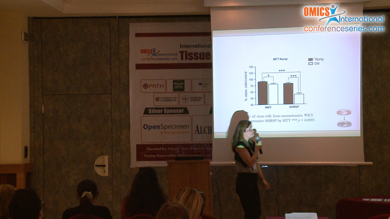
Mesenchymal stem cells (MSCs) hold tremendous potential for tissue regeneration. We hypothesized that the action of these cells is not only to repopulate the area of brain damage, but mainly to secrete neurotrophic and proliferative factors which could induce or stimulate the recovery of the damage. As we previously described, the model Stroke Prone Spontaneously Hypertensive Rat (SHRSP) exhibits a hippocampal damage, which can fully be recovered after transplantation of MSCs. Moreover, such cell repopulation occurred whereas apoptosis, superoxide and lipid peroxidation were reduced to normal levels, as verified in parallel with helpful Wistar Kyoto (WKY) controls (Calió et al; Free Rad Biol Med, 2014). The objective of this work is to investigate if transplanted MSCs could be involved in proliferation of neural cells by analysis of the levels of nitric oxide (NO) and expression of vascular endothelial growth factor (VEGF).
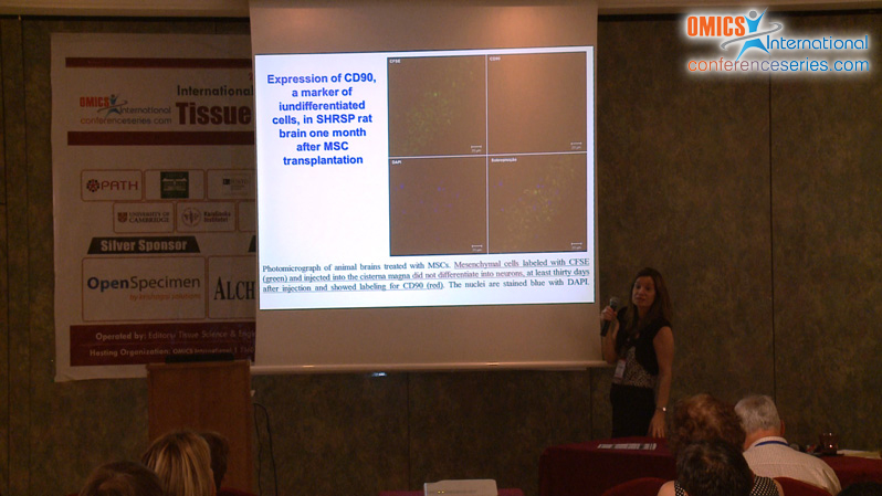
Methods We performed a comparison of the brains isolated from SHRSP treated or not with MSCs with those from normotensive Wistar Kyoto (WKY) controls by using quantitative RT-PCR, immunohistochemistry and biochemistry assays. MSCs were obtained from the femur and tibiae of 12-week-old WKY rats and characterized for the presence of mesenchymal surface antigens (CD90 and CD105) and the absence of hematopoietic markers (CD45, c-kit, and Sca1) by flow cytometry before the transplantation. 1x10⁶ MSC previously labeled with CFSE were injected into cistern magna through the atlanto-occipital membrane of 48-week-old SHRSP. The expression of VEGF was assayed both by Western blot and real-time (quantitative) RT-PCR analysis. β-Actin was used as a housekeeping gene. Nitric oxide (NO) was measured to evaluate its possible role in neural proliferation or endogenous brain cells regeneration after MSCs transplantation to the brain of stroke rat models. The quantification of generated NO was based on the gas-phase chemiluminescent reaction between NO and ozone; data were detected by the Nitric Oxide Analyzer (Sievers Instruments, Boulder, USA), a high-sensitive detector (~1 pmol) of NO in liquid samples.
Results An increase of almost threefold of VEGF expression was observed in the MSC-treated SHRSP group, thereby suggesting that transplanted stem cells have a proliferative potential by inducing proliferation of neural stem cells. Similar results were obtained in terms of elevation of generated NO detected by chemiluminescence. Thus, we suggest that both Vegf and NO can contribute to the recovery of hippocampal damage associated with oxidative stress and apoptosis in the spontaneously hypertensive stroke model SHRSP.
Conclusion: Our data suggests that MSCs could secrete or induce the same types of proliferation factors, such as Vegf and NO, which could stimulate the recovery of brain damage by endogenous stem cells. Thus, stem cell transplantation is a promising therapy, which could provide trophic support, helping promote survival, migration, and differentiation of endogenous precursor cells.
Supported by Fapesp, CNPq and FAP-Unifesp