Day 1 :
Keynote Forum
M Zavos
The Andrology Institute of America, USA
Keynote: The evolution and current status of sperm cryopreservation
Time : 9:45-10:10
Biography:
M Zavos received his BS in Biology-Chemistry in 1970, his MS in Biology-Physiology in 1972 and Education Specialist in Science (EdS) in 1976 from Emporia State University in Emporia, Kansas. He earned his PhD in Reproductive Physiology, Biochemistry and Statistics in 1978 from the University of Minnesota in the Twin Cities, Minnesota. He received the distinguished Alumnus Award and the Graduate Teaching Award from Emporia State University and the Student Leadership Award from the University of Minnesota. He has numerous scientific collaborations nationally and internationally and his publications have appeared in eight languages. He is a Member of the American Society for Reproductive Medicine (ASRM), the American Society of Andrology (ASA), and the European Society for Human Reproduction and Embryology (ESHRE), the Middle East Fertility Society (MEFS), the Japanese Fertility Society, the International Society of Cryobiology Sigma XI, Gamma Sigma Delta and a number of other Scientific and Professional Societies. He has served on a large number of committees for the International Society of Cryobiology, ASRM, MEFS, ESHRE and others.
Abstract:
Semen or sperm cryopreservation (commonly called sperm banking) is a procedure to preserve sperm cells. Semen can be used successfully indefinitely after cryopreservation which normally is kept at a very stable temperature of -196ºC (liquid Nitrogen). For human sperm, the longest reported successful storage which yielded a successful pregnancy is 22 years. The cryopreservation procedure can be used for sperm donation where the recipient wants the treatment in a different time or place, or as a means of preserving fertility for men undergoing vasectomy or other treatments that may compromise their fertility, such as chemotherapy, radiation therapy or surgery. Cryopreservation extends the availability of sperm for fertilization; however, the fertilizing potential of the frozen-thawed sperm is compromised because of alterations in the structure and physiology of the sperm cell. These alterations, characteristics of sperm capacitation, are present in the motile population and decrease sperm life-span, ability to interact with the female tract, and subsequent fertilizing ability. The etiology of such alterations may represent a combination of factors, such as inherited sensitivity of the sperm cell to withstand the cryopreservation process and the semen dilution. Although the former is difficult to address, approaches that make-up for the dilution of seminal fluid may be sought. The aim of this work is to review aspects of sperm cryopreservation paralleled by events of capacitation and evaluate the possible roles of sperm membrane cholesterol, reactive oxygen species, and seminal plasma as mediators of cryopreservation effects on sperm function. As far as the methods of cryopreservation of human sperm, there are three main conventional freezing techniques used in sperm cryopreservation: Slow freezing, rapid freezing and vitrification. The slow freezing technique which consists of progressive sperm cooling over a period of 2–4 hours in two or three steps, either manually or automatically using a semi programmable freezer; The rapid freezing technique which requires direct contact between the straws and the nitrogen vapors for 8–10 min and immersion in liquid nitrogen at −196°C and lastly, the vitrification method which is the newest procedure to be followed and which renders very high cooling and warming rates (over 20,000°C/min) and short contact with concentrated cryoprotective additives (less than 30 sec over −180°C) and which also offers a possibility to circumvent chilling injury and to decrease toxic and osmotic damage. All three of those methods are clinically employed and some of their technical aspects will be evaluated and compared along with their implications on the outcome of clinical results.
Keynote Forum
Heiko Zimmermann
Fraunhofer Institute for Biomedical Engineering, Germany
Keynote: Improved methods and procedures for pluripotent stem cell preservation, storage stability and validation
Time : 10:10-10:35
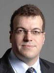
Biography:
Heiko Zimmermann is the Director and Head of Fraunhofer IBMT and Chair in Molecular and Cellular Biotechnology at the Saarland University and has been working as Physicist since 1997 in the field of cell biophysics. He coordinated the EU-project HYPERLAB in FP7 and was WP leader in several other EU-projects. Within EBiSC, he is Leader of WP 3 and is additionally coordinating the translational project DropTech® in FP7. He received the first permission for working with hESCs within the Fraunhofer Gesellschaft. He is the author of more than 70 peer-reviewed papers and book chapters. His research expertise covers cryobiology, cryotechnology and biopolymers for clinical scaffolds. He is inventor of more than 50 granted patent families from which more than 20 have been commercially licensed.
Abstract:
The project EBiSC aims to build up a European biobank for research grade human induced pluripotent stem cells (hIPSCs). The vision of EBiSC leads to the demand for upscaled production methods, these kinds of cells leading to the need for automated systems and procedures in stem cell processing and banking. An overview of existing state-of-the-art automation systems is given and the specifications for different applications are compared. Furthermore, modules and concepts for automated cell identification, pluripotency testing, and viability and functionality tests are drawn and results are shown. Scalable label-free analysis of pluripotent stem cells using quantitative life cell imaging and on-line image analysis is shown. A specialized system, the automated hanging drop technique (DropTech®) is shown. The DropTech system allows fully automated cultivation of hiPSCs on micro carrier using the hanging drop technology and enables applications like the automated Embryonic Stem cell Test (EST) for standardized embryo toxicity tests. The last part of the talk deals with the technology of cryopreservation, banking and validation frozen samples. The method of surface-based vitrification of pluripotent stem cells is introduced and the need for a completely closed cool chain is derived from experimental results. Solutions for automated industrial scale biobanking with closed cool chains and with minimal harmful thermal fluctuations are shown and the effect on functionality of cryopreserved cells compared to standard technology is shown. A method for non-invasive monitoring of re-crystallization and de-vitrification effects using Raman micro-spectroscopy is presented.
Keynote Forum
Igor Katkov
Belgorod National Research University, Russia
Keynote: Cryopreservation by vitrification: Basic thermodynamic principals, methods and devices
Time : 10:35-11:00

Biography:
Igor I Katkov is a trained Biophysicist with more than 30 years of experience in Cryobiology and Cryogenic Engineering. His research has been focused on the fundamental aspects of kinetic vitrification (K-VF) as well as on designing the practical system for K-VF KrioBlast™ in cooperation with V F Bolyukh from Ukraine. He is a Chief Scientific Officer of Celltronix, San Diego, USA and recently accepted a Professor-level position of the Head of the Laboratory of Amorphous State in the Belgorod National Research University, Russia.
Abstract:
Cryopreservation (CP) and subsequent long-term storage (cryobanking) are important parts of both life science research and related industries and technologies. As we have stated before, there are 5 basics ways of achieving long-term storage, all essentially lead to vitrification of cells, namely: Slow freezing (SF), Equilibrium vitrification (E-VF) with high concentration of vitrificants (“thickeners”) and relatively moderate speed of cooling and rewarming, Kinetic vitrification (K-VF) with very rapid rates of cooling and rewarming and low to none concentration of exogenous vitrificants, Freeze-drying (lyophilization), and Vacuum/air flow drying at temperatures above zero degree Celsius (xeropreservation), which up to now, is the mainstream of the majority of CP technologies. It however requires multi-step protocols, expensive programmable freezers and must be tuned to the particular types of cells, tissues and organs. In this presentation, we will focus on the kinetic vs. equilibrium vitrification. We will compare the mechanisms, analyze phase diagrams, emphasize the role of the Leidenfrost effect (LFE) and ways of reducing up to full eliminating LFE, discuss pros and cons of each methods and present information on basic equipment and accessories for E-KF and K-VF used in different field of biology, reproductive, regenerative and personalized medicine, drug screening, agriculture, conservation of endangered species, medicine and other related disciplines of sciences and industries. And, finally, we will introduce our (solution to hyper-fast cooling and present a short video clip of the novel hyper-fast scalable cooling devices KrioBlast™ and VitriPlunger™ and briefly discuss the promising results on K-VF of human sperm, embryonic stem cells and insulin-producing cells using the KrioBlas™ and VitriPlunger™ systems.
Keynote Forum
Charles F Mahl
GenLife Institute for Regenerative Medicine and Stem Cells, USA
Keynote: Prolotherapy the first choice in Regenerative Medicine
Time : 11.20-11:45

Biography:
Charles F Mahl is a graduate of Case Western Reserve University in Cleveland, Ohio. He has received his MD degree from the Rosalind Franklin School of Medicine in Chicago, Illinois. He did his Residency and Chief Residency in New York at Interfaith Hospital and Vitreo Retinal Fellowship at the University of Oregon. He has completed a Fellowship program in Anti-Aging and Regenerative Medicine by the American Academy of Anti-Aging Medicine, the Living Younger Preventive Aging Program and is certified in Age Management Medicine by The Cenegenics Education and Research Foundation. He is Regenestem trained in both PRP and adipose/bone marrow derived stem cells. He is Board Certified in Integrative Medicine (BCIM) and is a Certified Sports Trainer (CST) and Certified Sports Nutritionist (CSN). He is also a Member of the Advisory Board of Global Stem Cells and Regenestem.
Abstract:
Prolotherapy has been around for almost 80 years, yet is not well known in most musculoskeletal circles. Most orthopedic surgeons and neurosurgeons either have not ever heard of prolotherapy or think it does not work based on never having studied it in residency training programs, their personal bias or their general lack of knowledge about it. The author will explain his discovery of prolotherapy after some 30 years of suffering from pain and his experience and will introduce this fine medical art to the participants. Prolotherapy was the first procedure invented in Regenerative Medicine, followed by PRP-Platelet Rich Plasma injections and then Stem Cell Injections from the bone marrow and/or adipose tissue. Prolotherapy is a simple technique, much less invasive and less harmful as compared to steroid injections and high risk musculoskeletal surgery and often times, when performed by a skilled and experienced prolotherapist can alleviate the chronic pain one is suffering from. The author will explain the physiology of ligaments and tendons in response to prolotherapy and differentiate between 1st line, 2nd line and 3rd line regenerative medicine therapies. Prolotherapy is the first line of defense when it comes to regenerative medicine and should be recommended to patients prior to cortisone injections or invasive surgery. Elite athletes are already aware of its potential and utilize it when injured and or reinjured to heal their painful joints. It is time for prolotherapy to become main stream and no longer be considered alternative medicine. Author will describe the indications for prolotherapy, the techniques utilized and indications for this therapy. Prolotherapy is an extremely successful therapy in experienced hands and offers powerful pain relief and healing, especially to those who cannot afford PRP or Stem Cells and is therefore, a therapy that be part of all physicians training in third world countries and the like where large percentages of the population cannot afford or have no access to PRP or stem cells. For an indigent population, prolotherapy offers a very good treatment for healing and relief of musculoskeletal pain. Any physician doing stem cell therapy (musculoskeletal) should be well trained in prolotherapy prior to using the advanced therapies of performing platelet rich plasma injections (PRP) and bone marrow aspirates for stem cells and liposuctions for adipose derived stem cells. These procedures are very expensive as compared to prolotherapy. You need to have a major knowledge of injection techniques and know your anatomy-bones, ligaments, tendons and muscles in order to put the cells where they need to be. You need to be able to do these procedures via head, heart and hand (palpation) and via ultrasonic guidance and C-arm Fluoroscopy for more precision. Before rushing into doing stem cell therapies, prolotherapy should be tried because it is a much simpler and less invasive technique. Prolotherapy is currently being performed as a precursor to having PRP and stem cell therapy by competent regenerative medicine doctors. A discussion of what is prolotherapy and its uses and what it does is germane to this talk. It is important for physicians to know that often a simple treatment can be performed to accomplish the same result as long as one is experienced and knowledgeable as a prolotherapist.
- Biobanking | Cryopreservation| Vitrification | Biorepository & Biospecimen| Stem Cell | Regenerative medicine| Tissue engineering | Rejuvenation
Location: Plaza I
Chair
Panayiotis Zavos
The Andrology Institute of America, USA
Co-Chair
Debra Aub Webster
Cardinal Health Regulatory Sciences, USA
Session Introduction
Lourdes Cortes-Dericks
University of Hamburg, Germany
Title: Human mesenchymal stem cell-derived conditioned medium: Perspectives for therapeutic application in lung cancers
Time : 12:10-12:30

Biography:
Lourdes Cortes-Dericks completed her PhD in Biological Sciences from the University of Hamburg, Germany. Her dissertation was focused on the cellular and molecular mechanisms of the desensitization processes in guanylyl cyclase receptors. Her postdoctoral studies principally surround the tumor biology of lung cancers and identification of tumor biomarkers for tumor cancer stem cells to identify their roles in lung carcinogenesis. She is an active peer reviewer in numerous respected journals and an editorial board member.
Abstract:
Mesenchymal stem cells (MSCs) are adult stem cells having the capacity for self-renewal and differentiation. The current consensus of the therapeutic benefits of mesenchymal stem cells posits that these cells secret trophic factors that elicit paracrine actions for cancer therapy. These soluble factors may be collected in what has been known as the conditioned medium (CM). In the malignant setting, the biomolecules secreted by MSCs are thought to either support or inhibit tumor growth. This presentation will cover the anti-tumor effect of the human lung mesenchymal stem cell-derived conditioned medium (hlMSC-CM) in lung cancers. It will include the identification of the different biomolecules contained in the hlMSC-CM and its anti-tumor effects in malignant mesothelioma cell lines. It will highlight the efficacy of hlMSC-CM as compared to twice the IC50 of cisplatin in eliminating the chemo resistant, sphere-forming mesothelioma cells. Other regenerative capacities MSC-CM from our and other studies such as the attenuation of cigarette smoke-induced injury in lung fibroblasts, in vivo and in vitro will also be mentioned to underscore its potential in improving tissue regeneration.
In the clinical setting, the use of cell-free MSC-derived conditioned medium may facilitate a practical approach compared with stem cell-based therapy as the former is easier to prepare, preserve and transport to appropriate clinics, and may also present less complications relative to issues on cell transplantation. Despite the promising results of the beneficial effects of MSC-derived conditioned media, standardization of MSC cultivation, collection and preservation of the CM is still necessary.
Xu Han
CryoCrate LLC and University of Missouri, USA
Title: Life in Nano Ice : Application of cryocrate C80EZ medium for cell and tissue cryopreservation
Time : 12:30-12:50

Biography:
Xu Han has over 10 years of experience in the field for Cryopreservation. He is the Top Reviewer of the journal of Cryobiology, and a winner of NIH SBIR, Coulter Translational Partnership Program Award, and MU Faculty Innovation Award. He has endeavored in improving cryopreservation methods and devices. His expertise is also in the fields of Calorimetry, Numerical Simulation, Thermodynamics and Mechanics.
Abstract:
Traditional cryopreservation approaches are technically problematic in either producing large ice crystals (typically on um scale) associated with equilibrium freezing process or involving extremely high concentrations (40-60% v/v) of permeating cryoprotectants to achieve vitrification. Both also require the use of liquid nitrogen and expensive facilities for long-term storage or banking. With the support of NIH SBIR, Coulter Translational Partnership Program, and University of Missouri (MU) Fast-track Award, CryoCrate (a MU spin-off) has developed an innovative cryopreservation medium, trademarked as C80EZ, and opened the door for cryopreservation with both nano scale ice formation and long-term storage in regular deep freezers at approximately -80ºC. The base medium of C80EZ enables long-term storage of cells at -80ºC by preventing recrystallization and the efficiency has been demonstrated by previous publications, e.g. Sci Report 2016 (nature.com). The complete C80EZ medium enables unique nano scale ice formation and the efficiency tests have been performed on cell or tissue types that survive poorly from any traditional cryopreservation method, e.g. Human corneas, primary neurons, porcine and rodent spermatozoa, and cell types that cannot be stored in deep freezers for extended periods of time, e.g. prophetical blood mononuclear cells and mouse embryos. Significant improvement of cell post-thaw viabilities and functionalities has been observed for all above cell and tissues types. For E. coli competent cells (routinely stored in deep freezers), our preliminary data showed that the nano ice technology can remarkably increase post-thaw transformation efficiency. Currently, the C80EZ medium doesn’t improve survivability from cryopreservation for cell types with large volume of intracellular lipid components, e.g. porcine and fish embryos. The C80EZ medium is now commercially available, and the technology is patent pending (PCT filed) and exclusively licensed from MU to CryoCrate.
Jedediah Lewis
Organ Preservation Alliance, USA
Title: The promise of organ banking and large-scale support for cryopreservation advances
Time : 12:50-13:10

Biography:
Dr. Jedediah Lewis currently serves as Chief executive officer at Organ Preservation Alliance. Dr. Jedediah Lewis holds degree from Harvard law school and also work as a research assistant in Harvard law school and Emory university school of medicine.
Abstract:
A growing research effort aims to achieve the cryopreservation of viable large tissues and organs for both transplantation and research, with the potential to save or improve the lives of millions of patients worldwide. Recent years have seen multiple targeted federal grant pipelines for complex tissue cryobanking that have funded dozens of labs across the U.S., as well as roundtables at the White House and on Capitol Hill, an NSF-funded technology road mapping process, and other meetings with U.S. science agencies and stakeholder organizations to generate support for organ and tissue banking progress. At two global Organ Banking Summits, held at sites including Stanford School of Medicine and Harvard Medical School, leading cryopreservation labs have mapped out the remaining “sub-challenges” to cryo bank whole organs and large tissues in a viable state. The American Society of Transplantation recently launched a community of practice that aims to support advances toward the cryopreservation of whole organs, limbs, and an array of other biological systems. These developments offer numerous opportunities for the biobanking community, as they can enable both new applications of bio banked tissue, new cold chain solutions for cells and tissues, and new support for biobanking advances.
Yulian Zhao
Mayo Clinic, USA
Title: Reproductive tissue banking for young boys of pre-reproductive age
Time : 14:10-14:30

Biography:
Yulian Zhao is an Associate Professor of Gynecology and Obstetrics and Director of Assisted Reproductive Technology Laboratory at the Department of Gyn/Ob, the Johns Hopkins University School of Medicine. She is an Officer, Board Member, Committee Member and Active Member in a number of professional organizations and societies in the field of Reproductive Medicine. She frequently acts as an ad hoc reviewer for over 12 professional journals and has been a recipient of several academic awards. She has authored over 50 peer-reviewed articles, review articles and book chapters as well as delivered over 50 presentations at the professional conferences and society events nationally and internationally.
Abstract:
Loss of fertility can significantly compromise quality of life. For populations of reproductive age, fertility preservation strategies have been well established and widely used, but most of the technologies are not options for pediatric age group, because of their sexual immaturity. Majority of such cases would be cancer patients. One of the medical late effects of cancer treatment is infertility or severely compromised fertility. Fertility preservation is of great importance to psychological well-being and to the quality of life for these patients. Fertility preservation for this population has become an issue of attention in recent decades. Strategies for long-term preservation and storage of reproductive tissue for this particular population are of key importance for scientific research and reproductive medicine. This emerging field encompasses cryopreservation of reproductive cells including testicular cell suspensions and testicular tissue. There are 3 potential strategies under active investigation. The first one is to cryopreserve testicular tissue or cell suspension before cancer therapy. Once the patient is cured from cancer and ready to begin a family, the tissue or cells could be thawed and re-implanted into the patient's own testes to continue full maturation. The second option is in vitro differentiation of testicular tissue. Spermatozoa may be generated from spermatogonial stem cells in vitro until they can be used for in vitro fertilization or be re-implanted into the testes. The third one is that immature cryopreserved testicular tissue may be grafted into another organ or tissue of the patient. These fertility restoration techniques have been successfully applied in several animal models and are considered to be very promising for future clinical applications in human.
Jill Davies
Oxford University, UK
Title: Fertility cryopreservation in Oxford, UK
Time : 14:30-14:50

Biography:
Jill Davies graduated from Coventry University in 1987 with a degree in Applied Biology. After university studies, she worked for cardiothoracic surgeons Mr. Donald Ross & Sir Magdi Yacoub at the National Heart Hospital in London and that she opened the heart valve bank at the Oxford University Hospital in Oxford, England in 1990. The Oxford bank supplies cardiovascular tissue, corneas for transplant and research and brains & spinal cords for research. It is now also an Oxford Research Center Bio bank. She is also an executive member of the BATB, member of AATB, SLTB, and Society for Cryobiology.
Abstract:
Late effects of cancer treatment may cause premature infertility in children and adolescent cancer survivors. Oxford applied for UK license for cryopreservation of ovarian and immature testicular tissue (ITT) for patients requiring gonadotoxic treatment. This required validation with animal/human studies, development of protocols, information, staff training, establishing costs/funding source. Oxford recruited first set of patients in 2013. Patients eligible for treatment intent is curative, cytoxicity high risk, age <40 years (females) and boys pre-pubertal. Patients consented prior to day of surgery. Ovarian tissue from 145 females, age 10 months – 39 years cryopreserved (14% with benign diagnosis), testicular tissue (ITT) from 31 boys, (0.9–13.6 years). Referrals from oncologists/hematologists and patients, most patients travel to Oxford for surgery scheduled to coincide with other interventions (minimizing risk of anesthesia). Right ovary procured using laparoscopic oophorectomy, wedge testicular biopsy procured via midline incision. Tissue bank technician collects tissue and dissects within cleanroom, producing ovarian cortical strips (1x1x5mm) containing primordial follicles or testicular segments (2x2x2 mm) containing spermatagonial stem cells. Histological evaluation reviews viability and presence of metastatic cells. Immature oocytes collected for in vitro maturation (IVM) and vitrified. Cryopreservation of ovarian tissue requires ethylene glycol, sucrose, human albumin serum, slow controlled freezing (manual seeding at 8ËšC) and vapor phase nitrogen storage. DMSO is used for ITT. Tissue is stored until the patient is cured and confirmed infertile. 86 live births following ovarian tissue transplantation was recorded. A live birth in ITT is successful in animal studies only. Patients with hematological malignancy will use vitrified oocytes (gametes are free from residual disease). For other females and ITT patients, tissue will be matured in vitro (IVM) to develop primordial follicles/spermatogenesis for use in conventional ART. Oxford University researchers currently are optimizing IVM techniques (three dimensional/organ culture studies). All details were registered in OTCP database. Service evaluation (interviews with patients/parents) confirms patients/parents understood risks/potential for fertility restoration. Setting up the Oxford service has been challenging due to evolving UK legislation. Referrals are from all UK centers.
Mohamed A Zayed
Washington University School of Medicine, USA
Title: Peripheral artery and blood biobanking can lead to scientific collaboration and discovery
Time : 14:50-15:10

Biography:
Mohamed A Zayed is Surgeon-Scientist at the Department of Surgery, Section of Vascular Surgery, and Washington University School of Medicine, USA. He has completed his Medical training at Stanford University and earned his Doctoral degree in Pharmacology at the University of North Carolina at Chapel Hill. He has previously served as the Chief Medical Officer for a software start-up company and has published over 30 research articles in reputable journals. His current clinical and research interests focus on the influences of diabetes on peripheral arterial disease.
Abstract:
Objectives: Over 3 million Americans have advanced peripheral arterial occlusive disease leading to significant patient morbidity and mortality. The lack of well-preserved human peripheral arterial tissue substrate has limited scientific exploration of this disease process and development of impactful-targeted molecular therapies. To address this, we developed an integrative biobanking strategy to collect peripheral arterial tissue specimens from patients undergoing vascular surgery.
Methods: Over 29 months, we harvested vascular specimens from consenting patients undergoing open arterial endarterectomy and revascularization procedures. All patients were enrolled in an IRB approved protocol. A biobank infrastructure was developed to manage logistics, funding, collection, and real-time processing of harvested arterial tissue.
Results: 486 patients were enrolled in the vascular surgery biobank prior to their index operation. Forty-two (42) clinical variables were evaluated for each patient during the perioperative period. Vascular specimens were successfully collected for 72.3% (349) of patients who enrolled in the biobank. The majority of specimens collected were retrieved from the peripheral arterial system (38.7% carotid artery, 9.5% anterior or posterior tibial arteries, 24.4% femoral or popliteal arteries). For all patients, blood samples were collected and processed to provide serum (85.5%) and plasma (86.1%). Each arterial specimen was sub-divided into maximally and minimally diseased portions to facilitate intra- and inter-patient biochemical and molecular analyses. Over the study period 8 collaborations (in 4 different departments at 2 universities) were fostered to provide 82 tissue specimens and 98 blood samples.
Conclusions: An integrative biobanking approach in vascular surgery patients is feasible and provides a highly unique peripheral arterial substrate for molecular and biochemical analyses. Biobanking management and daily operations requires a dedicated team approach to insure proper patient consenting, specimen collection, subsequent experimental analysis and meaningful scientific collaborations.
Dalip Sethi
Cesca Therapeutics Inc., USA
Title: Clinical applications of autologous bone marrow derived cells
Time : 15:10-15:30

Biography:
Dalip Sethi currently serves as Director of Clinical Research at Cesca Therapeutics Inc., a public company engaged in the research, development, and commercialization of cellular therapies and delivery systems for use in regenerative medicine. He holds a PhD in Biotechnology from Institute of Genomics & Integrative Biology, New Delhi, and a Master’s in Organic Chemistry from Hansraj College, Delhi University, India, and conducted Post-doctoral studies at Thomas Jefferson University, School of Medicine. He is currently engaged in the development of point-of-care devices, methods and diagnostics for use in autologous cell therapy in the treatment of cardiovascular and orthopedic diseases.
Abstract:
Cardiovascular diseases are a major burden on healthcare in modern society. Diseases, such as Critical Limb Ischemia (CLI), are debilitating. Many of the cardiovascular ischemic disease patients have limited surgical or medical options. Regeneration of vascular system is an attractive treatment strategy and is actively pursued in various preclinical and clinical settings. One of the options in regenerative medicine is the use of autologous bone-marrow concentrate (aBMC) containing stem and progenitor cells. Autologous bone-marrow concentrate (aBMC) is derived from the bone marrow aspirate (BMA) by density centrifugation and can be delivered either intra-muscularly (IM) or intra-coronary in the affected region. The aBMC consists of a) an acellular fraction comprised of autologous plasma and the cytokines and b) cellular fraction which is a source of (i) proangiogenic cells such as hematopoietic stem cells, mesenchymal progenitor cells, and endothelial progenitor cells; (ii) other cells of immune system at different levels of maturity and multi-potency. The acellular and cellular components participate in tissue repair and regeneration and have made aBMC an attractive source of cells and cytokines for therapeutic angiogenesis in the treatment of ischemic diseases.
Raffaella Fabbri
University of Bologna, Italy
Title: Ovarian function recovery after transplantation of ovarian tissue cryopreserved and stored for long-term

Biography:
Raffaella Fabbri has been working as a Biologist in the Human Reproductive Medicine Unit, S Orsola-Malpighi Hospital of University of Bologna in Italy since 1977. In 2001, she received a national and international worldwide patent about method and solution for cryopreserving oocytes. In 2006, she obtained the United States Patent US 7,011,937 B2 for method and solutions for cryopreserving oocytes. She has her expertise in cryopreservation of human mature and immature oocytes, cryopreservation of embryos and blastocysts, cryopreservation of human ovarian tissue in vitro maturation of ovarian follicles, co-cultures of human granulosa cells and sperms, fertilization and embryonal development. She has been involved in many scientific activities which are well-documented by 335 articles published in Italian and foreign journals, participated in 134 meeting as speaker and 58 as Auditor.
Abstract:
Statement of the Problem: Ovarian tissue cryopreservation represents a valid strategy to preserve ovarian function in cancer patients with a high risk of premature ovarian failure due to chemo/radiotherapy. The ovarian tissue remains frozen for very long period of time (the request of tissue replanting usually occurs after at least five years from the end of therapies and this period may dragging on further in the case of diseases that require prolonged treatments or in the case of pediatric patients). The purpose of this study is to evaluate the morphology and functional activity of cryopreserved ovarian tissue stored for 18 years after thawing and transplantation.
Methodology & Theoretical Orientation: Ovarian tissue of a 29 year old patient suffering from Hodgkin Lymphoma was cryopreserved at our Centre before starting anticancer treatment. 18 years after storage; her ovarian tissue was evaluated by light microscopy, transmission electron microscopy, TUNEL assay and LIVE/DEAD viability/cytotoxicity test and then heterotopically transplanted in two subcutaneous pockets of patient. Follicle development was evaluated by ultrasound examination on the graft sites.
Findings: Ovarian tissue showed a good morphology, no apoptosis signs, sub-cellular integrity of follicles and interstitial edema foci. The LIVE/DEAD assay performed on stromal cells, isolated from cryopreserved tissue, showed viable cells (>97%) after 2 and 7 days of culture. The patient had the first menstruation five months after transplantation and to date (20 months from the graft), she is regularly menstruating every 30-40 days. Follicular development is monthly evidenced by a bulge palpable beneath the skin in the graft sites.
Conclusion & Significance: This study provides evidence that the storage time does not impact on tissue quality and on tissue ability to resume the ovarian function after replanting. These results give hope especially to cancer girls, whose tissues could remain cryopreserved for a very long time.
Achour Radhouane
El-Manar University, Tunisia
Title: Premature ovarian failure diagnosis and management
Time : 15:30-15:50

Biography:
ACHOUR Radhouane is associate professor at faculty of medicine of Tunis-Tunisia; He has published many basic and clinical articles in relation to gynecology and obstetrics. , his research interests include Rare Diseases in gynecology and prenatal diagnosis.
He serves as associate professor, Emergency Department of Gynecology and Obstetrics in maternity and neonatology center Tunis ; Faculty of Medicine of Tunis- El Manar University of Tunis-Tunisia. He also serves as member of the editorial team for: Asian Pacific Journal of Reproduction, the Global Journal of Rare Diseases, Journal of Neonatal Biology, Current pediatric research, Obstetrics and Gynecology: Open access, Pediatrics and Health Research and Member of the Science Advisory Board.
Abstract:
Introduction: Premature ovarian failure (POF) is defined as menopausal levels of follicle stimulating hormone (FSH≥40 IU/l) associated with more than 4 months of secondary amenorrhea occurring before the age of 40. It affects approximately 1% of women and the underlying an etiology remains very complex and heterogeneous. Recently Welt suggested changing the term POF to POI, which stands for premature ovarian insufficiency, because this better reflects the longitudinal progression towards the final menstrual cycle.
Objective: The objective of this study was to investigate different etiologic mechanisms of premature ovarian failure.
Material & Methods: A retrospective study of 30 cases of early menopause reported in our service for period of 10 years from January 2000 to December 2009. We will center this work to draw up an epidemiologic profile of our population, to specify the various circumstances of discovery, to analyze the various mechanisms etiopathologic and finally to discuss the therapeutic possibilities.
Results: The incidence of premature ovarian failure is around 1 to 3%. This pathology occurs in young women who often wish to become pregnant. Two mechanisms could be involved: Initial follicle depletion and follicle dysfunction; however in some cases mixed mechanisms are involved initially. Our study includes 30 patients. Genetic anomalies were found in 3 cases (10%). Anti-ovarian antibodies were found in 18 cases (60%). Ovarian biopsy done in 20 patients confirms immunological mechanism of ovarian failure in 16 cases (70%) with autoimmune ovaritis. In 9 cases idiopathic ovarian failure was observed.
Discussion: The POF etiology in women with normal karyotype remains poorly understood. However, more and more genes involved in the normal follicular and ovarian development are becoming known, which are also likely to be involved in this disease, based on the data obtained from animal models. In such context, the first study presented herein was conducted to evaluate whether the ovarian biopsy, when compared with data provided by pelvic ultrasound (US), could be a reliable tool to individualize patients with an ovarian phenotype suggestive of a particular gene mutation, therefore, suggesting the usefulness of laparoscopy and ovarian histology in orientating the search for the possible genetic etiologies of the POF syndrome. In our experience, when women had no follicle at US, histological examination did not detect follicles in most of our patients. At the contrary, only 56% patients with normal size ovaries with the presence of follicles suggested at US, displayed follicles when histological examination of an ovarian biopsy was performed. Consequently, it can be assumed that, US is not predictive of the presence of follicular structures within the ovary.
Conclusion: The proportion of idiopathic premature ovarian failure tends to decrease owing to the better recognition of genetic and immunological anomalies. Finally the use of the ovarian biopsy still remains interesting in our hands to help us to better understand the process of ovarian dysfunction and is mandatory in patients for whom anomalies of genes involved either in follicular growth or maturation are found.
Brian M Mehling
Blue Horizon International, USA
Title: Mesenchymal stem cells in clinical applications
Time : 16:30-16:50

Biography:
Brian M Mehling is a practicing American Orthopedic Trauma Surgeon, Researcher, and Philanthropist. He started his path in Medicine through Undergraduate study at Harvard University, obtaining Bachelor of Arts and Master of Science degrees in Biochemistry from Ohio State University. Completing his degree of Medicine at Wright State University School of Medicine, he received Post-graduate education through residencies and fellowships at St. Joseph’s Hospital in Paterson, NJ and the Graduate Hospital in Philadelphia, PA, while pursuing a PhD in Chemistry. He operates his own practice, Mehling Orthopedics, in both West Islip, NY and Hackensack, NJ.
Abstract:
Mesenchymal stem cells (MSCs), multipotent adult stem cells feature the potential to regenerate tissue damage and inhibit inflammation. Our research studies designed to measure the safety and efficacy of intra-venous, intra-articular, and intra-thecal stem cell therapies. Retrospective Chart Review study of umbilical cord blood (UCB) stem cell therapy for spinal cord injury showed that significantly higher proportion of patients in the stem cell therapy group showed improved function in pain and temperature sensation, lower limb muscle strength, bladder and gastrointestinal function compared to the traditional therapy group. UCB stem cell therapy of patients with Sequelae of cerebro-vascular hemorrhage and CVA (Stroke) Sequelae showed significant improvement in mobility and muscle strength of upper and lower extremities, improvement of neurological function. In the period from 2015 to 2016, 207 subjects with musculo-skeletal conditions underwent the therapy with their own stromal vascular fraction cells. Analysis of subjects’ pain sensation and mobility showed the following: 10 days after the therapy, pain sensation decreased by 26.8%. Three months after therapy, the number of subjects with a decrease in pain sensation increased to 50.0%, and six months later, this number was 52.5%. Analysis of subjects’ mobility showed the following: 10 days after the therapy, mobility was improved by 26.0%. Three months after therapy, the number of subjects with improved mobility increased to 43.8%, and six months later this number was 47.4%. Statistical analysis showed that the decrease in subjects’ pain sensation and improvement of subjects’ mobility three and six months after the therapy was statistically significant.
Kay Ohlendieck
Maynooth University, Ireland
Title: Proteomic profiling of tissue degeneration and regeneration in muscular dystrophy

Biography:
Kay Ohlendieck has an Undergraduate degree in Biology from the University of Konstanz, Germany (1985), a PhD in Biochemistry from University College Cork, Ireland (1989) and a DSc in Muscle Biology from University College Dublin, Ireland (2011). He has worked as a Post-doctoral Associate at the University of Iowa, Iowa City and at the State University of New York, Stony Brook, as well as a Lecturer in the Department of Pharmacology, University College Dublin (1995-2001). Since 2002, he is Chair of Biology at the National University of Ireland, Maynooth, and his research focuses on skeletal muscle proteomics
Abstract:
Statement of the Problem: The X-linked neuromuscular disorder Duchenne muscular dystrophy is caused by primary abnormalities in the dystrophin Dmd gene and is associated with progressive skeletal muscle wasting, sterile inflammation and reactive myofibrosis. The purpose of this study was to analyze tissue degeneration and regeneration in different muscle subtypes that exhibit varying degrees of pathophysiological susceptibility to fiber necrosis.
Methodology & Experimental Approach: A comparative mass spectrometry-based proteomic analysis was carried out to determine distinct alterations in various skeletal muscles from the dystrophic mdx and mdx-4cv animal models. This included the characterization of muscle proteins in mildly versus severely affected skeletal muscles, as well as the label-free mass spectrometric evaluation of proteome-wide changes following fiber regeneration. The independent verification of proteomic findings was carried out by comparative immunoblotting surveys with isoform-specific antibodies to key muscle protein markers and immune-fluorescence microscopy.
Findings: In this study, the complexity of changes in the skeletal muscle proteome during tissue degeneration and regeneration could be established. Muscle-associated proteins belonging to the contractile apparatus, excitation-contraction coupling mechanism, metabolic pathways and the cellular stress response were shown to undergo changes in their abundance and/or isoform expression pattern. Contractile proteins, molecular chaperones, mitochondrial enzymes, cytoskeletal proteins and components of the extracellular matrix exhibited differential changes in moderately versus severely dystrophic fiber populations. During regeneration cycles, the reversal of a variety of protein changes was established by comparative proteomics.
Conclusion & Significance: The newly identified proteomic markers of tissue degeneration and regeneration might represent suitable biomarker candidates to advance our general understanding of the molecular pathogenesis of muscular dystrophy, as well as being useful for improving the diagnosis, prognosis and therapy monitoring of dystrophinopathies
Debra Aub Webster
Cardinal Health Regulatory Sciences, USA
Title: Regenerative medicine–from promise to patient
Time : 17:10-17:30

Biography:
Debra Aub Webster has over 20 years of experience in Pharmaceutical Research and the Regulatory Environment. She started her regulatory career with the US FDA as Reviewing Toxicologist/Pharmacologist. As a Principal Scientist in Regulatory Affairs and Product Development with Cardinal Health Regulatory Sciences, she is the Project Lead for biologic and regenerative medicine product development programs. In this capacity, she provides guidance on clinical, nonclinical, and regulatory aspects of strategic product development, author regulatory documents, and acts as the regulatory representative for sponsors in interactions with FDA.
Abstract:
The expansion in the use of human cells and tissues from their functions in practice of medicine, for repair or replacement, to their use as a manufactured product intended to treat an indication or condition has led to increased regulatory oversight by FDA. Under the authority of Section 361 of the PHS Act, FDA introduced a comprehensive regulatory program in 1997 for human cells, tissues, and cellular and tissue-based products (HCT/Ps). This consolidated regulatory approach is tiered and risk-based to allow for less regulatory evaluation of products determined to present a minimal risk to patient safety. In 2005, this regulatory program was implemented in rules codified under 21 CFR Section 1271. Under these rules, some HCT/Ps are defined as biological products, requiring market clearance. The potential regulatory pathways for HCT/P-derived technologies is often complex, and understanding these regulatory pathways and the definitions surrounding them is critical to successful product development. Navigating the regulatory path, from scientific discovery to initiation of clinical investigations is challenging. These challenges include determination of the appropriate regulatory pathway and understanding the requirements regarding demonstration of safety and efficacy and identification of appropriate quality attributes regarding product safety, quality, and potency. FDA has issued numerous guidance documents and offers several expedited approval pathways in order to bring effective discoveries safely forward, to benefit patients. Successful strategies for achieving this goal include identifying critical information gaps with respect to the appropriate regulatory pathway; informed clinical, non-clinical, and quality development plans; maintaining a Target Product Profile; and successful interaction with the appropriate review division within FDA.
Robin C Muise-Helmericks
Medical University of South Carolina, USA
Title: pGlcNAc nanofibers derived from a marine polymer stimulate regenerative wound repair via activation of a TLR4/type-I IFN pathway in combination with an integrin/Akt1 dependent pathway
Time : 17:30-17:50

Biography:
Robin C Muise-Helmericks is currently an Associate Professor in the Department of Regenerative Medicine and Cell Biology, Member of the Hollings Cancer Center and Adjunct Associate Professor in the Departments of Oral Health at the Medical University of South Carolina.
Abstract:
The structural balance between elastin, collagen and other extracellular matrix components is absent in adult wound repair resulting in misaligned and excessive collagen deposition that contributes to scar formation. Our findings show that a marine derived pGlcNAc nanofiber stimulates tensile strength and elasticity of wounded skin so that it is equal to that of uninjured skin. Nanofibers promote cellular alignment, secretion of aligned collagen fibers and increased tropoelastin expression. Nanofiber-dependent reductions in scar formation are, in part, due to activation of an integrin/Akt1-dependent pathway. In addition to the activation of Akt1, our deep sequencing results suggest a role for a TLR4/type-I interferon (IFNαβ) pathway in the nanofiber-driven anti-fibrotic phenotype. Here we show that pGlcNAc nanofibers specifically stimulate type-I IFN (IFNαβ) expression via activation of Toll-like receptor 4 (TLR4), in the absence of TLR4-induced NFκB-dependent inflammatory responses in normal human fibroblasts and endothelial cells. TLR4 null animals fail to respond to nanofiber treatment, which can be rescued by addition of IFNα2A to the wound bed. Indeed, inhibition of type-I IFN activity using a blocking antibody against the type-I IFN receptor inhibits nanofiber induced tissue repair. Our findings support a model where nanofiber stimulation of TLR4 preferentially favors the IFNαβ response over an inflammatory response mediated through MyD88. As inflammatory responses lead to increased TGF-β production, increases in myofibroblast production and fibrotic, disorganized collagen deposition. Our findings suggest that reductions in inflammatory cell recruitment or activation results in reduced scar formation and increased tensile strength and elasticity of healed wounds. The marked difference in collagen deposition and increased elastogenesis in nanofiber treated wounds suggests that the nanofibers are specifically stimulating a more regenerative type of tissue repair.
Joel I Osorio
Westhill University School of Medicine, Mexico
Title: Therapeutic effects of bioquantine® in volunteer patients diagnosed with Cerebral palsy, Spinal cord injury, Wet amd, Sci, Hypothyroidism, Atypical pyoderma gangrenosum, and Breast cancer
Time : 17:50-18:10

Biography:
Dr. Osorio brings over 20 years of experience in medical practice. Served in capacities in private and public practices, as hospital staff physician and as emergency health services coordinator for a variety of private and public institutions throughout Mexico and managed a blood bank from 2009- 2012. Earned MD degrees at Westhill University and the National Autonomous University of Mexico as a Medico Cirujano. In addition, has diplomas in aesthetic medicine from the Autonomous University of Guadalajara, as an Advance Fellow by the American Board of Anti-Aging and Regenerative Medicine, visiting scholar at University of North Carolina at Chapel Hill in dermatology. Fellow of Stem Cell Medicine by the American Academy of Anti-Aging Medicine and University of South Florida, and currently completing additional masters work in metabolic and nutrition sciences at University of South Florida and Master in Health Sciences Aged Services, Victoria University at Melbourne Australia. A member of the Round table of the ReGeNeRaTe laboratories Mexico’s Committee, a DNAge-Lab Company, working on the stem cells field since 2007, and since 2011 a member of the International Cellular Medicine Society. Chief Medical Officer & Co-Founder at Astrum Salud Móvil in Mexico City. PRP certified practitioner in the aesthetic and regenerative field since 2011 by GyCo.
Abstract:
We have previously demonstrated that co-electroporation of Xenopus laevis frog oocytes with normal cells and cancerous cell lines induces the expression of pluripotency markers, and in experimental murine model studies that Bioquantine® extract (purified from intra- and extra-oocyte liquid phases of electroporated oocytes) showed potential as a treatment for a wide range of conditions Squint, Spinal Cord Injury (SCI), Cerebral Palsy and skin wrinkling. The current study observed beneficial changes with Bioquantine® administration in a small group of patients with a variety of degenerative disorders. Pluripotent stem cells have therapeutic and regenerative potential in clinical situations including dermatological and CNS disorders even cancer. One method of reprogramming somatic cells into pluripotent stem cells is to expose them to extracts prepared from Xenopus laevis oocytes. We showed previously that co-electroporation of Xenopus laevis frog oocytes; with normal cells and cancerous cells lines, induces expression of markers of pluripotency. We also observed therapeutic effects of treatment with a purified extract (Bioquantine) of intra- and extra-oocyte liquid phases derived from electroporated X. laevis oocytes, on experimentally induced pathologies including murine models of melanoma, traumatic brain injury, and experimental skin wrinkling induced by squalene-monohydroperoxide (Paylian et al, 2016). The positive human findings for Squint, Spinal Cord Injury, Cerebral Palsy and wrinkling with the results from previous animal studies with experimental models of traumatic brain injury, skin wrinkling, and melanoma,respectively (Paylian et al,2016). Because of ethical reasons, legal restrictions, and a limited numbers of volunteers, we were able to treat only a very small number of patients. These results indicate that Bioquantine® may be safe and well tolerated for use in humans, and deserves further study in a range of degenerative disorders. We propose that the mechanism of action of Bioquantine® in these various diseases derives from its unique pharmacology and combinatorial reprogramming properties. In conclusion, these preliminary findings suggest that Bioquantine is safe and well tolerated and contains ubiquitous therapeutic activity including restoration Squint (extropia),Cerebral Palsy, dermatologic healing, spinal cord injury.
Nan Xiao
University of the Pacific, USA
Title: Glial cell derived neurotrophic factor promotes dental pulp stem cell migration
Time : 18:10-18:30

Biography:
Nan Xiao is an Assistant Professor of the Department of Biomedical Sciences, Arthur A. Dugoni School of Dentistry, University of Pacific. She is a member of the American Dental Education Association (ADEA) and International Association of Dental Research (IADR). She serves as editor for multiple scientific journal. She also works intensively to promote the dental academic career pathways for dental students. Her academic interest include research in application dental pulp stem cells mediated tissue regeneration and integration the pedagogic innovations in dental education.
Abstract:
Preserving the vitality of the teeth is critical in maintaining the function and esthetics during dental treatment. Dental pulp stem cells (DPSCs) are mesenchymal cells that are demonstrated to possess stem cell properties, such as self-renewal, proliferation and pluripotency. DPSCs can be obtained through non-invasive procedure from the dental pulp and become potential resources for tissue regeneration. Neurotrophic factors are known to promote survival and growth of neurons. In the present study, we examined the expression of the glial cell derived neurotrophic factors (GDNF) family ligands and receptors, and characterized the intracellular localization of them in DPSCs. GDNF family ligands increased the migration of the DPSCs. In addition, we found the AKT and MAPK pathways are downstream of GDNF in regulating the DPSC wound healing and migration. Our results indicate that neurotrophic factors play a role in dental pulp regeneration and may be potential novel therapies for post pulpotomy treatment in adult teeth.
- Biobanking | Cryopreservation| Vitrification | Biorepository & Biospecimen| Stemcell | Regenerative medicine| Tissue engineering | Rejuvenation
Location: Plaza I
Chair
Heiko Zimmermann
Fraunhofer Institute for Biomedical Engineering, Germany
Co-Chair
Ken yaegaki
Nippon Dental University School of Life Dentistry, Japan
Session Introduction
Aijun Wan
University of California, USA
Title: Engineering stem cells and biomaterials to treat birth defects before birth
Time : 14:20-14:40

Biography:
Abstract:
Elvira Famulari
Molecular Biotechnology Center, Italy
Title: Cell therapy for Crilger Najjar Type I syndrome
Time : 18:00-18:15
Biography:
Elvira Famulari is pursuing Ph.D in Molecular Biotechnology Center from University of Turin Italy.
Abstract:
Wael Abo Elkheir
Military Medical Academy, Egypt
Title: Regenerative effect of autologous mesenchymal stem cell injection on cartilage defects
Time : 17:40-18:00

Biography:
Abstract:
Hala Gabr
Cairo University, Egypt
Title: Role of bone-marrow derived stem cells in liver regeneration: a multicenter clinical trial
Time : 17:20-17:40

Biography:
Abstract:
Jin-Ye Wang
Shanghai Jiao Tong University, China
Title: Natural Polymer, Zein for Tissue Regeneration
Time : 17:00-17:20

Biography:
Abstract:
Shrikant L Kulkarni
Kulkarni Clinic, India
Title: Hydro-pressure therapy in chronic kidney diseases
Time : 16:40-17:00

Biography:
Abstract:
Lin-Hwa Wan
National Cheng Kung University, Taiwan
Title: Application of ultrasonography in the assessment of overhand movement
Time : 16:20-16:40

Biography:
Abstract:
Yong Jian Geng
University of Texas, USA
Title: Regeneration and repair of post-infarct cardiovascular tissues with rejuvenated stem cells
Time : 15:40-16:00

Biography:
Abstract:
Stephen S. Lin
California Institute for Regenerative Medicine, USA
Title: Initiatives to advance stem cell science and medicine at California’s $3 billion stem cell agency
Time : 15:20-15:40

Biography:
Abstract:
Seungil Ro
University of Nevada, USA
Title: Smooth Muscle Genome/Transcriptome Browser: Offering genome-wide references and expression profiles of transcripts expressed in intestinal primary cells and tissues
Time : 15:00-15:20

Biography:
Abstract:
Joanne Mullarkey
University of Bradford, UK
Title: Developing and setting up a tissue donation after death service in the UK: A research nurse’s personal perspective
Time : 10:15-10:35

Biography:
Abstract:
Vittorio Sebastiano
Stanford School of Medicine, USA
Title: Pluripotent Cells: A tool to model development and to deliver definitive cure for pediatric diseases
Biography:
Abstract:
Sita Somara
Wake Forest Institute for Regenerative Medicine, USA
Title: Clinical translation of tissue-engineered medical products (TEMP) - Journey from bench to bed-side
Time : 14:00-14:20

Biography:
Abstract:
Bo Feng
University of Hong Kong, China
Title: CRISPR/Cas9-Mediated knock-in of large DNA in human embryonic stem cells and somatic cells
Time : 12:35-12:55

Biography:
Abstract:
Pao Chi Liao
National Cheng Kung University, Taiwan
Title: Biomarker discovery using mass spectrometry-based proteomics/metabolomics and biospecimens
Time : 12:15-12:35

Biography:
Abstract:
Won-Gun Koh
Yonsei University, Korea
Title: Hydrogel micro pattern-incorporated nanofibers for biomedical application
Time : 11:55-12:15

Biography:
Abstract:
Nidhi Bhutani
Stanford school of medicine, USA
Title: ChallengesBiologics to enhance mesenchymal stem cell expansion and storage of developing a Biobank in Oxford, UK
Time : 11:35-11:55

Biography:
Abstract:
Jill Davies
Oxford University, UK
Title: Challenges of developing a biobank in Oxford, UK
Time : 10:35-10:55

Biography:
Abstract:
Jan Huebinger
Max-Planck Institute of Molecular Physiology, Germany
Title: Cryopreservation of living cells using electron microscopy fixation methods


